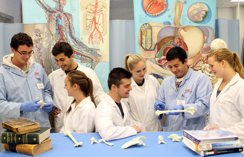
“I think a short video is worth a million words,” says William Albabish. Exploring the worth of videos and photos in teaching human anatomy is the common purpose of new PhD projects by Albabish and Sean McWatt, both in the Department of Human Health and Nutritional Sciences (HHNS).
As the first-ever doctoral candidates working in U of G’s human anatomy teaching lab, they are developing videos and high-resolution photographs of body organs and systems. Their projects are intended to yield new teaching tools for undergrad students learning about anatomy from whole-body dissections and prosections in the lab’s third-year course. The videos and photos being developed this year will follow the curriculum as students dissect cadavers from week to week in the year-long course.
Prof. Lorraine Jadeski, HHNS, expects these learning resources will help the lab run more efficiently. By viewing the tools before a lab session, students can prime themselves for that day’s dissection. Viewed afterward, the videos and photos will help students review.
Also available during classes, the resources will provide on-the-spot refreshers and a handy alternative to posing questions of a teaching assistant or course instructor. “The goal is to develop learning tools to help make students self-sufficient,” says Jadeski.
Adds Albabish: “They can rely less on the TAs and more on themselves.”
Hands-on dissection is still the primary learning approach in the lab, says Prof. Lawrence Spriet, chair of HHNS. “This is not going to change what we do with the students. We still believe doing hands-on dissections yourself is powerful. This is one more tool for students to help study and learn where things are.”
Offering different tools also helps accommodate varied learning styles. “Not everyone learns the same,” says Spriet. Through dissection and prosection, textbooks and multimedia, “Any additional resources you can provide will enhance the chances that you’re going to reach all the students in your class.”
Videos and photos are also intended to spark students’ curiosity and deepen their understanding of organs and systems. “My project is to give more students access to anatomy,” says Albabish.
He took anatomy in the third and fourth years of his human kinetics program. “It was a big life changer. I fell in love with the study of anatomy,” he says. “That’s where I grew my love of teaching.”
For his master’s degree with Jadeski, he filmed a partial dissection of the upper back. Besides creating seven 10-minute video modules, he used 2,300 photos to create a two-and-a-half-minute time-lapse video of the dissection and added a voice-over explaining the procedure. He’s now creating a series of video modules covering a full-body dissection; the videos will accompany lab sessions, one by one.
Discussing his project at a teaching and learning conference this year, he had a number of Toronto-area colleges asking how they might gain access to anatomy videos.
Besides teaching almost 400 Guelph students in human kinetics and biomedical science each year, the human anatomy program teaches thousands of visiting health-care professionals and college and high school students.
Also a human kinetics grad, McWatt began taking high-quality photographs in the lab for his master’s project with Jadeski, who is compiling an anatomy atlas for the course.
As with the videos, the detailed photos will be used in teaching modules covering a year’s dissection. “We will introduce the modules to the class and see how that alters their engagement and approach to learning,” says McWatt. “We’re not only developing tools to use in the lab for students, but we’re also trying to gauge how effective they are.”
Each week, those anatomy students spend three hours in dissections and two hours examining dissected parts, or prosections. McWatt says these new tools will allow students to continue their learning outside of the lab and will be targeted to Guelph’s course approach and curriculum.
Both his father and his grandfather did veterinary studies at Guelph. Sean had considered a similar path but was attracted by human kinetics and biomechanics. “It’s science and biology, but it’s also artistic. I found a good balance.”
Both PhD students are co-supervised by HHNS professor Genevieve Newton, who studies undergraduate learning in the department.
For more information on U of G’s human anatomy program, view a promotional video produced for the department’s Giving for Life fundraising program.